Transperineal prostate fusion biopsy: how it is done and what to expect
If you have a raised PSA or an MRI showing a suspicious area, you may be offered a prostate biopsy. Here I explain when it is considered, how it is done (through the perineum, under general anaesthesia or deep sedation and fusing micro-ultrasound with MRI), how to prepare and what to expect afterwards.
Reviewed by Dr. Mario Domínguez Esteban, urologist (medical licence no. 392860199). Head of the Uro-Oncology Section, Marqués de Valdecilla University Hospital.Last reviewed: · Next review: Oct 2027
When is it considered?
A biopsy is the test that confirms or rules out prostate cancer. Small tissue samples are taken with a fine needle, and a pathologist examines them under the microscope. It is mainly considered in these situations:
The MRI shows a suspicious area (PI-RADS 4 or 5) or an equivocal one (PI-RADS 3), and the rest of the data support investigating it.
Your PSA is still raised when repeated, and the risk calculator estimates a risk that justifies checking.
You have already had a negative biopsy, but the suspicion remains.
You have low-risk cancer on active surveillance and it needs to be checked that it has not changed.
It is not always needed. If the MRI shows no suspicious area and your risk is low, a biopsy can sometimes be avoided and you continue with check-ups. The full pathway is in High PSA: what it means and what to do. If your PSA is high because of benign prostate enlargement and you have urinary symptoms, in HoLEP I explain one of the treatment options.
First, the MRI: what PI-RADS means
Before a biopsy, the usual step is an MRI scan of the prostate. Each area seen is scored on the PI-RADS scale, from 1 to 5, according to how likely it is that there is a cancer needing treatment:
PI-RADS 1-2
Unlikely.
PI-RADS 3
Equivocal. Whether to do a biopsy depends on other data, such as PSA density (the ratio between PSA and prostate size) and the risk calculator.
PI-RADS 4-5
Likely or highly likely. A biopsy is usually considered.
MRI does not replace a biopsy. It shows whether one is advisable and where to take the samples.
On what artificial intelligence adds (and what it does not) when interpreting prostate MRI, you can read AI in prostate MRI.
How it is done
The biopsy is done under general anaesthesia or deep sedation, so you are not aware of the procedure. This way you do not move and the image fusion is accurate. At the end, local anaesthetic is given so that you have less discomfort afterwards.
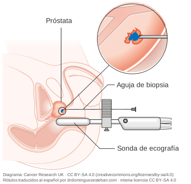
High-resolution ultrasound. A high-resolution micro-ultrasound probe (ExactVu) is placed in the rectum and shows live images of the prostate.
Fusion with the MRI. The MRI images are overlaid on the ultrasound images. This shows at all times where the suspicious area is.
Needle through the perineum. The needle goes in through the skin of the perineum, the area between the scrotum and the anus, not through the rectum.
Samples. Targeted samples are taken from the suspicious area and, usually, also some from the rest of the prostate.
For years, biopsies were almost always taken through the rectum (transrectal route). Today the European guidelines recommend the transperineal route, for two reasons:
Lower risk of infection. The needle does not pass through the rectum. In a review of 165 studies, serious infection (sepsis) occurred in 0.1% of transperineal biopsies and in 0.9% of transrectal ones.
It reaches the whole prostate well. Including the front part, which is harder to reach by the transrectal route.
If you have no risk factors, antibiotics are often not needed. Your doctor will assess whether you need them.
Before: preparation and medication
Bring the report and images of your MRI, and your PSA results.
Tell us if you take anticoagulants or antiplatelet drugs (for example, aspirin or clopidogrel). We will tell you whether and when to stop them. Do not stop them on your own.
As it is done under anaesthesia, you will need to fast beforehand and have the pre-operative tests you are given.
If you have a fever or symptoms of a urine infection in the days before, let us know: it may need to be postponed.
Come with someone: you should not drive on the day of an anaesthetic.
Afterwards: discomfort and when to seek help
In the following days it is common to notice blood in the urine or semen. Blood in the semen can last several weeks. You may also notice mild discomfort in the perineum.
If you have a fever or cannot pass urine, seek urgent medical attention.
Results
The pathologist examines the samples. This takes a few days (usually between 5 and 7), and the results are discussed at your appointment.
If there is cancer, the report states how far the cells differ from normal ones. This is the grade, expressed as the Gleason score (from 6 to 10) and its equivalent, the ISUP grade group (from 1 to 5). The higher the grade, the more likely the cancer is to grow and spread.
With the grade, the PSA, the MRI and the examination, the risk is estimated, and you and your urologist decide the next steps: follow-up with new tests, further investigations or treatment. Low-risk cancer does not always need immediate treatment: monitoring it (active surveillance) is often enough.
Who performs it
Dr. Mario Domínguez Esteban is a urologist and Head of the Uro-Oncology Section at Marqués de Valdecilla University Hospital. He performs fusion biopsy with high-resolution micro-ultrasound (ExactVu), which he introduced in his private practice years ago, and reads prostate MRI (EAU-certified training course).
He has studied how to combine PHI and MRI to decide who needs a biopsy (BJUI Compass 2025, in Sources).
On video
Video in Spanish · 1 min 37 s · Spanish subtitles
Prostate biopsy: how it is done today
MRI first; if there is a suspicious area, a fusion biopsy targeted at that area and by the transperineal route, with a lower risk of infection.
AI-generated avatar video; script reviewed by Dr. Domínguez Esteban.
Show transcript (English translation)
Have you been offered a prostate biopsy? Today it is done differently from a few years ago.
In the past, samples were taken from all over the prostate, without really knowing where to look. And almost always through the rectum.
Today, the usual approach is to start with an MRI scan. The MRI shows whether there is a suspicious area. If there is not, the biopsy can sometimes be avoided.
If there is a suspicious area, a fusion biopsy is performed. The MRI images are overlaid on the live ultrasound. This way the needle is guided to that area. Usually some samples are also taken from the rest of the prostate.
Where does the needle go in? The European guidelines recommend the transperineal route. The needle goes in through the skin between the scrotum and the anus, not through the rectum. This means a lower risk of infection. And, if you have no risk factors, antibiotics are often not needed.
It is often done under general anaesthetic or deep sedation. That way you do not move, and the image fusion is precise. It is also more comfortable for you. Local anaesthetic is also given so that you have less discomfort afterwards.
In the following days you may notice blood in your urine or semen. This is common. If you have a fever or cannot urinate, seek urgent medical attention.
At drdominguezesteban.com you will find the raised PSA pathway, with this step explained.
When to seek help
Seek urgent medical attention if after the biopsy you have a fever, cannot pass urine, pass clots in your urine or feel faint.
Book a priority appointment with your doctor if the blood in your urine is not decreasing over the days.
Not during the procedure, because it is done under general anaesthesia or deep sedation. At the end, local anaesthetic is given to reduce discomfort. In the following days you may notice mild discomfort in the perineum.
Why general anaesthesia or deep sedation?
This way you do not move, the image fusion is accurate and the procedure is more comfortable for you. The European guidelines note that a biopsy can also be done under local anaesthesia alone.
Do I need to prepare?
Yes. Before the procedure you will need to use an enema. Also, as it is done under anaesthesia, you will need to fast beforehand and have the pre-operative tests you are given. If you take anticoagulants or antiplatelet drugs, tell us: we will tell you whether and when to stop them. Do not stop them on your own. The details are in Before: preparation and medication.
Will I go home the same day?
Yes. It is an outpatient procedure: afterwards you spend a while in the recovery room and go home the same day. Come with someone, because you should not drive on the day of an anaesthetic.
Transperineal or transrectal?
The European guidelines recommend the transperineal route because it has a lower risk of infection: the needle goes in through the skin of the perineum and does not pass through the rectum. It also reaches the front of the prostate well.
Do I need antibiotics?
With the transperineal route, if you have no risk factors, they are often not needed. Your doctor will assess whether you need them in your case.
Does PI-RADS 3 always mean a biopsy?
No. PI-RADS 3 is an equivocal area. The decision is made together with PSA density, the risk calculator and the rest of your case. Sometimes repeating the check-ups is enough.
When will I get the results?
Examining the samples takes a few days (usually between 5 and 7). The results are discussed at your appointment.
Is blood in the semen or urine normal?
Yes, it is common in the following days. If there are clots, you cannot pass urine, you have a fever or you feel faint, seek urgent medical attention.
How long does the biopsy take?
The procedure itself takes about 30 minutes. You must arrive fasting; with anaesthesia (general or deep sedation) and about 60 minutes in recovery, expect to spend about two hours at the centre.
When can I resume normal activity, exercise or sex?
Take it easy for the next few days and avoid strenuous exercise and cycling. You can resume sex when you feel well; blood in the semen can last several weeks and is not harmful to your partner.
What does micro-ultrasound (ExactVu) add?
It provides real-time images of the prostate at higher resolution than conventional ultrasound. Fused with the MRI, it helps guide the needle to the suspicious area.
What is cognitive fusion biopsy?
It is when the urologist guides the needle by looking at the MRI and the ultrasound separately, without overlaying them on one screen. In software fusion, the two images are overlaid.
Is it covered by my insurance?
Coverage depends on each policy; Urología Mínimamente Invasiva (UMI) can tell you.
And in the public health system?
In the public health system, your GP refers you to the Urology department if they consider it necessary. The technique and type of anaesthesia depend on each hospital's protocols.
If you would like me to assess your case, Where I see patients explains how to book an appointment, both in the public and in the private sector.